Figure 6.
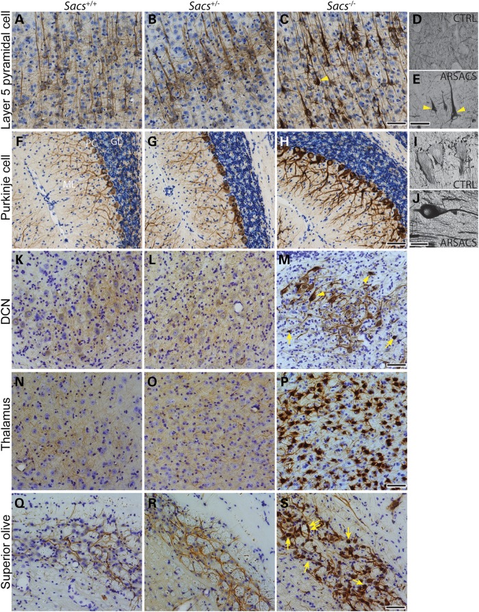
NF accumulations in Sacs−/− and ARSACS neurons. (A–C), (F–H) and (K–S) NFH immunohistochemistry on sagittal brain sections of 180-day-old mice and on brain sections from a human control autopsy case (D and I) and one ARSACS case (E and J). NF accumulations are present in Sacs−/− and ARSACS neuronal populations, such as layer 5 pyramidal cells (A–E), Purkinje cells (F–J), neurons of the deep cerebellar nuclei (DCN) (K–M), thalamic neurons (N–P) and superior olive neurons (Q–S). Nuclear displacement to the periphery is shown in pyramidal cells and neurons of deep cerebellar nuclei with NF accumulations (arrowheads in C, E and M). NF containing swellings are seen in neurons of deep cerebellar nuclei and superior olive (arrows in M and L) (scale bar = 100 µm in C, H, M, P and L and 50 µm in E and J).

