Figure 5.
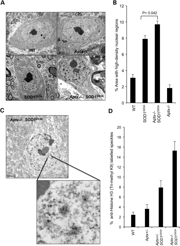
Increased nuclear invaginations and elevated Histone H3 K9 trimethylation in Aptx−/− SOD1G93A neurons. (A) Representative transmission electron microscopy (TEM) images of motor neurons prepared for ultrastructural analysis from 3-month-old WT, Aptx−/−, SOD1G93A and Aptx−/− SOD1G93A mice. Scale bar = 5 μm. (b) The average nuclear area occupied by high-density nuclear regions was quantified from ∼70 neurons obtained from three independent litters. Data are the average ± SEM. (C) Representative TEM images of H3 K9 trimethylation revealed by immunogold labelling of motor neurons from 3-month-old Aptx−/− SOD1G93A mouse. Scale bar = 2 μm (top) and 0.2 μm (bottom). (D) Average nuclear area occupied by the anti-Histone H3 (trimethyl K9)-positive dense chromatin within the indicated genetic backgrounds was quantified from 30 neurons obtained from three independent litters. Data are the average ± SEM.

