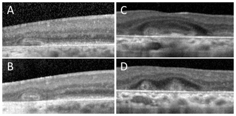
Figure 2.

Evolution of outer retinal tubulation (ORT) over time. Eye-tracked spectral-domain optical coherence tomography (SD-OCT) scans for varying conditions at baseline (top row) and follow-up (bottom row). A, B, SD-OCT scans of an eye with choroideremia at baseline and 35 months later, showing the development of ORT from the gradual invagination of outer retinal structures, at the junction of intact and atrophic retina. C,D, SD-OCT scans of an eye with Stargardt disease, showing long and ovoid ORT structures evolving into smaller round structures, over a period of 5 months.
