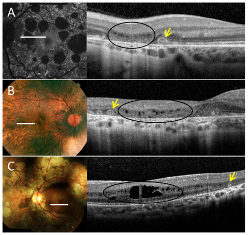
Figure 4.

Outer retinal tubulation (ORT) in near proximity to sites of cystoid macular edema (CME) in various degenerative disorders. A, Fundus autofluorescence (left) and corresponding spectral-domain optical coherence tomography (SD-OCT) (right) of an eye with pattern dystrophy, which highlights the distinguishing features of ORT (arrows), such as hyperreflective borders and positioning within the outer nuclear layer, from the findings of CME (black ovals). B-C, Fundus photographs (left) and SD-OCTs (right) of eyes with B, choroideremia, and C, gyrate atrophy.
