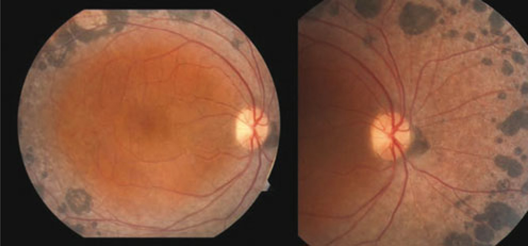
Fig. 1.
The fundus photograph of the right eye from patient 1. Extensive subretinal nummular pigmentation, located mostly in the mid-periphery along the vascular arcades, is visible. There is no indication of peripheral or central retinoschisis or maculopathy. No additional abnormal findings were found within the vascular arcades

