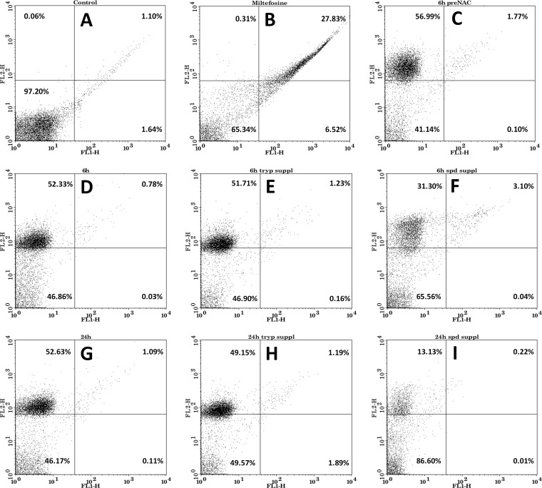
FIG 6.
Analysis of the mode of cell death of Leishmania promastigotes. (A) Promastigotes without any treatment. (B) Promastigotes treated with miltefosine (50 μM) for 24 h. (C) Promastigotes pretreated with N-acetylcysteine (20 mM) before hypericin (18 μM) treatment for 6 h. (D) Promastigotes treated with hypericin (18 μM) for 6 h. (E) Promastigotes supplemented with trypanothione (0.5 μM) before hypericin (18 μM) treatment for 6 h. (F) Promastigotes supplemented with spermidine (100 μM) before hypericin (18 μM) treatment for 6 h. (G) Promastigotes treated with hypericin (18 μM) for 24 h. (H) Promastigotes supplemented with trypanothione (0.5 μM) before hypericin (18 μM) treatment for 24 h. (I) Promastigotes supplemented with spermidine (100 μM) before hypericin (18 μM) treatment for 24 h. Samples were stained with annexin V-FITC and propidium iodide and were analyzed by using flow cytometry. Promastigotes were shown to undergo necrosis after treatment with hypericin.

