
FIG 2.
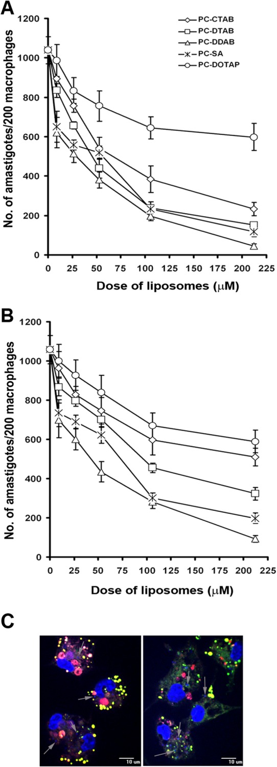
(A and B) Effects of graded concentrations of various cationic liposomes on L. donovani amastigote proliferation within murine peritoneal macrophages, using the AG83 (A) and GE1F8R (B) strains. Values are means ± standard errors of three independent experiments. (C) Uptake of PC-SA (left) and PC-DDAB (right) liposomes inside infected macrophages. Peritoneal macrophages isolated from BALB/c mice were infected with L. donovani promastigotes for 3 h and subsequently treated with fluorescent PC-SA or PC-DDAB liposomes. Macrophage nuclei are stained with DAPI (large blue areas). Intact liposomes are visible close to the amastigote nuclei, colocalizing green and red fluorescence and appearing yellow. Arrows, L. donovani amastigotes (small blue areas) inside macrophages. Magnification, ×40. Data are representative of two independent experiments.