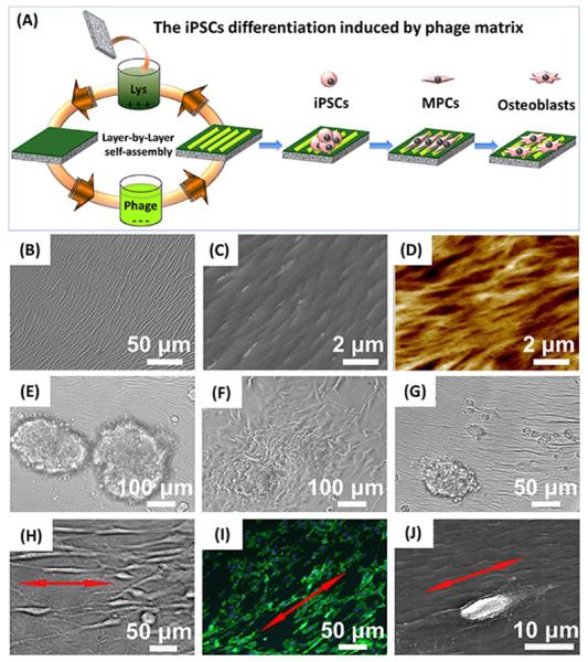
Figure 2.
Fabrication of phage-assembled matrix and the interaction between the phage matrix and iPSCs. The phage-assembled matrix with an ordered surface nanotopography was generated by a layer-by-layer self-assembly method, where the glass substrate was alternately immersed into the cationic poly-l-lysine solution and anionic phage nanofiber solution to achieve the electrostatic self-assembly of phage matrix, and phage layer was the last layer deposited (A). The resultant phage matrix showed specific ridge/groove nanotopography (B, bright-field image; C, scanning electron microscopy (SEM) image; D, AFM image). The resident iPSCs-derived EBs showed a quick response to the phage matrix and exhibited a significant morphological alteration over culture time (E, 6 h; F: 24 h; G, 48 h; H-J, 2 weeks). The fibroblast-like cells were first present at the cluster edge (E), and their number increased after 24 h (F). Some fibroblast-like cells were detached from the cluster edge and became slightly aligned along the phage bundles after 48 h (G). We also discovered that these fibroblast-like cells were significantly elongated and aligned along the long axis of the phage bundles constituting the phage matrix (H–J, red arrows showed the direction of cell elongation along phage bundles). Cell nuclei and F-actin were stained by DAPI (blue) and FITC-labeled phalloidin (green), respectively.

