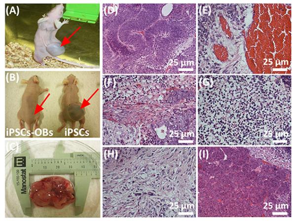
Figure 4.
Teratoma evaluation of iPSCs-derived osteoblasts (iPSCs-OBs). The animal test demonstrated that the iPSCs-Ob did not induce obvious teratoma formation in vivo, whereas pure iPSCs generated a large teratoma (around 3 cm in length) one month after both iPSCs-Ob and iPSCs were separately injected into the nude mice (A, injection of iPSCs into live nude mouse to generate a large visible teratoma; B, comparison of nude mice injected with iPSCs-Ob and iPSCs; C, teratoma excised from the animal). Histological analysis further demonstrated that the formed teratoma contained three embryonic germ layers including ectoderm (D and E), mesoderm (F, G and H) and endoderm (I). D, Immature neural tissue, ectoderm; E, Blood vessel tissue, ectoderm; F, Muscle-like tissue, mesoderm; G, Cartilage tissue, mesoderm; H, Bone tissue, mesoderm, I, Intestinal track tissue, endoderm. The red arrows in (A) and (B) showed the injection site.

