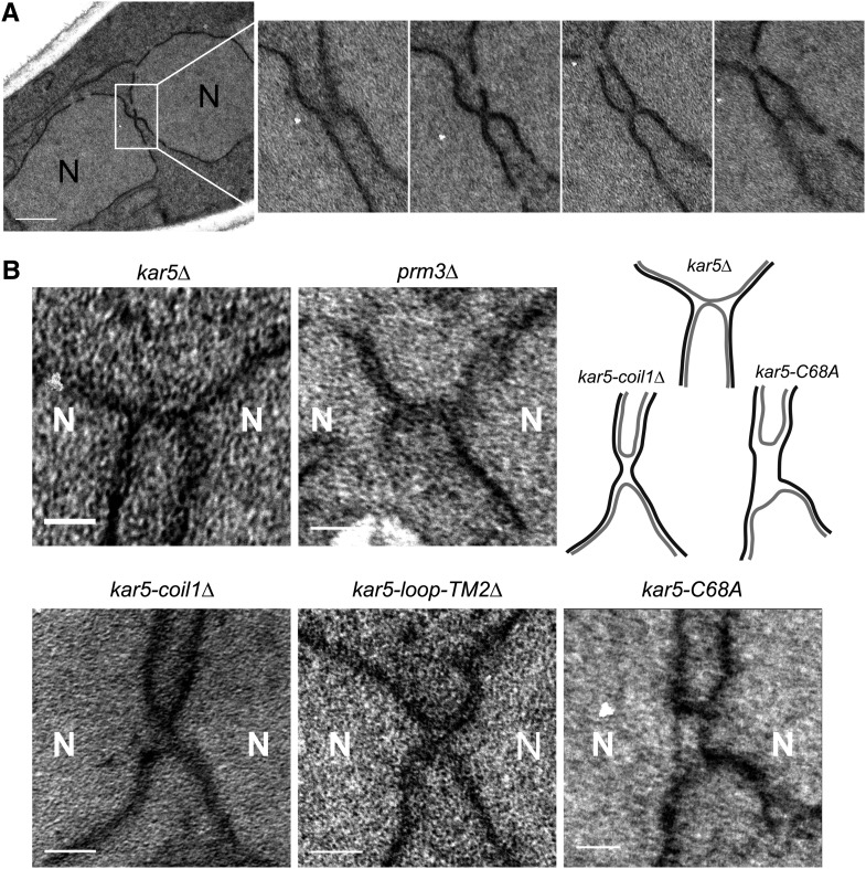
Figure 6.
Electron microscopy of kar5 mutants that may mediate distinct functions. (A) Four representative serial sections (~80 nm thick) of a kar5-loop-TM2Δ zygote (see the section Materials and Methods for details). Note the tight membrane bridge observed in the second serial section. N = nucleus. Scale bar, 500 nm. (B) Representative membrane bridges from other kar5 mutants. Each zygote pair is kar5Δ (MS7670) × kar5Δ (MS7673) with the indicated CEN plasmid in both parents. kar5Δ indicates that the strain is carrying an empty CEN vector (pMR1868). For comparison with a mutant arrested at outer membrane fusion, we also imaged prm3Δ (MS7590) × prm3Δ (MS7591). An interpretive cartoon of the kar5Δ, kar5-coil1Δ and kar5-C68A is shown. Outer nuclear envelopes are in gray, inner nuclear envelopes are in black. All zygotes are oriented with the two nuclei along the horizontal axis. Scale bars, 100 nm.

