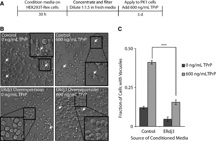
Figure 4. Secreted ERdj3 attenuates vacuole formation induced by toxic prion protein in mammalian cells.

- A Schematic illustrating the treatment of PK1 cells with ERdj3-conditioned media. HEK293T-Rex (Control) and HEK293T-Rex cells stably overexpressing tet-inducible ERdj3WT on poly-D-lysine-coated plates were treated with dox (1 μg/ml) overnight to induce ERdj3WT expression. The media was replaced with fresh OptiMEM I + 5% FBS, which was conditioned for 30 h, sterile-filtered, concentrated 5× against a 3 kD MWCO membrane, and diluted 1:1.5 with fresh OptiMEM I + 5% FBS. PK1 cells were plated in OptiMEM I + 5% FBS and after 24 h were treated with conditioned media and TPrP (600 ng/ml) for 3 days.
- B Representative images under 136× magnification of PK1 cells incubated with control conditioned media collected from HEK293T-Rex or conditioned media prepared from HEK293T-Rex cells overexpressing ERdj3WT in the presence or the absence of TPrP (600 ng/ml), as described in (A). Arrows indicate representative cells containing vacuoles. Insets show 544× magnification of a typical region of the image.
- C Quantification of vacuole formation from images as shown in (B). Equal area images comprising ∼1,200 cells per well were counted for each sample. Counting of cells and of cells with identifiable vacuoles was done using the Fiji image processing package (Schindelin et al, 2012). ***P-value < 0.001 (n = 3); mean ± SEM.
