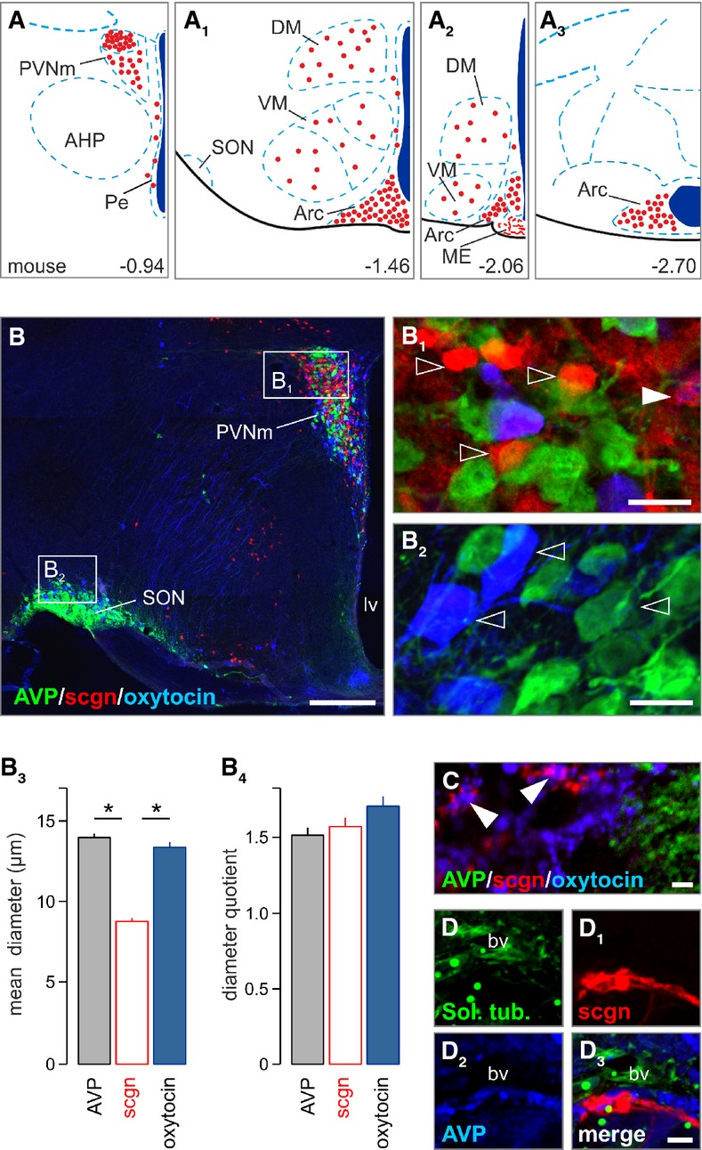
Figure 1. Secretagogin locus in the paraventricular nucleus of the hypothalamus.

- A–A3 Secretagogin+ neurons populate the paraventricular, dorsolateral, ventromedial, periventricular and arcuate nuclei of the mouse hypothalamus (Paxinos & Franklin, 2001). Red circles denote the localization of neuronal perikarya and their relative densities. AHP, anterior hypothalamus; Arc, arcuate nucleus; DM, dorsomedial nucleus; ME, median eminence; Pe, periventricular nucleus; PVN, paraventricular nucleus, PVNm, magnocellular part; SON, supraoptic nucleus; VM, ventromedial nucleus.
- B–B2 Largely non-overlapping distribution of AVP+, oxytocin+ and secretagogin+ (sgcn+) neurons in the magnocellular PVN. The mouse supraoptic nucleus (SON) harbored vasopressin+ and oxytocin+ but not secretagogin+ neurons. Open arrowheads pinpoint single-labeled neurons. Solid arrowhead denotes AVP/secretagogin dual-labeling. lv, lateral ventricle; PVNm, magnocellular part of the paraventricular nucleus; scgn, secretagogin.
- B3, B4 Secretagogin+ neurons had smaller somatic diameters than AVP+ or oxytocin+ neurons yet without a difference in their diameter quotient, a measure of ovoid profiles (*P < 0.05, Student’s t-test).
- C–D3 Terminal-like profiles in the posterior pituitary (arrowheads in C) suggesting that secretagogin can co-exist, even if infrequently with oxytocin. Solanum tuberosum lectin (Sol. tub) was used to identify blood vessels.
Data information: Scale bars: 100 μm (B), 10 μm (B1, B2), 2 μm (C–D3)
