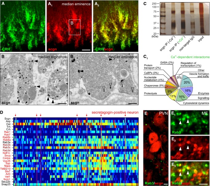
Figure 5. Secretagogin is a Ca2+ sensor protein.
- A–A2 Secretagogin co-existed in the majority of CRH+ nerve endings in the median eminence (ME). Open rectangle denotes the general location of (B, B1).
- B, B1 Large axon terminals in the median eminence were immunopositive for secretagogin with silver-intensified immunogold particles (open arrowheads) associated with axonal membrane and dense-core vesicles (see Fig 1D for quantitative data). Solid arrowheads denote silver deposit particles proximal to the plasmalemma.
- C Immunoprecipitation using an anti-secretagogin antibody in Ca2+-free and Ca2+-loaded conditions was subtractively used to decipher the Ca2+-dependent protein interactions. Silver-stained gel is shown.
- C1 Ontology classification of the 99 protein hits based on primary function assignments. Unbiased MALDI-TOF proteomics was used to identify interacting proteins. The most abundant hits (Supplementary Table S3 is referred to for details on individual proteins) are proteins implicated in vesicle fusion, trafficking, transport and formation and the regulation of vesicle exocytosis. “Other” refers to a group of proteins without known function.
- D Single-cell transcriptomics was used to validate the above proteome data by clustering mRNA transcripts encoding proteins that underpin vesicular release processes (red color labels highest mRNA abundance, whereas dark blue color indicates the lack of mRNA expression).
- E–E3 Rab3, a family of vesicular fusion and transport proteins (Schluter et al, 2006), was found co-localized with secretagogin (arrowheads) in the median eminence. Neuronal soma in the PVN lacked appreciable co-localization. Note that neither our MALDI-TOF analysis nor our histochemical probing allowed the precise identification of individual Rab3C-E family members.
Data information: Scale bars: 10 μm (A1, E3), 7 μm (E), 500 nm (B), 150 nm (B1).

