Figure 3.
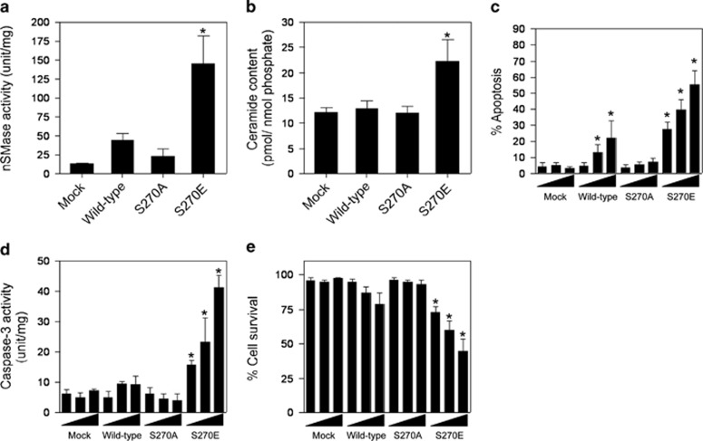
Induction of ceramide generation and apoptosis by the overexpression of mutant S270E in zebrafish ZE cells. (a) The nSMase activities of nSMase1 transfectants. ZE cells were transiently transfected with three different doses (1, 2, or 3 μg) of mock, FLAG-tagged wild-type, S270A, or S270E SMase1 vector DNA constructs and cultured for 24 h. nSMase activity was measured using C6-NBD-sphingomyelin as a substrate. Column 1, mock; column 2, wild-type; column 3, S270A; column 4, S270E. (b) The ceramide content in nSMase1 transfectants. Cellular lipids were extracted from the nSMase1 transfectants, and the levels of ceramide were quantified using a diacylglycerol kinase assay after thin layer chromatography separation. Column 1, mock; column 2, wild-type; column 3, S270A; column 4, S270E. (c) Apoptosis in nSMase1-transfected cells. ZE cells were transfected with three different doses (1, 2, or 3 μg) of mock, FLAG-tagged wild-type, S270A, or S270E SMase1 vector DNA constructs, cultured for 24 h, and stained with DAPI to quantify apoptosis. (d) Caspase-3 activity in nSMase1 transfectants. Caspase-3 activation was assessed by measuring Ac-DEVD-MCA hydrolysis. (e) The viability of nSMase1-transfected cells, as determined using Trypan Blue dye exclusion. (f) Apoptotic cells in the nSMase1 transfectants after heat shock. ZE cells were transfected with three different doses (1, 2, or 3 μg) of nSMase1 constructs per dish and cultured for 24 h. After heat shock at 38 °C for 1 h, the cells were incubated at 25 °C for up to 23 h, and all apoptotic cells were identified using DAPI staining. Each value represents the mean of three independent experiments, and the error bars represent the S.D.s. *P<0.01 versus mock

