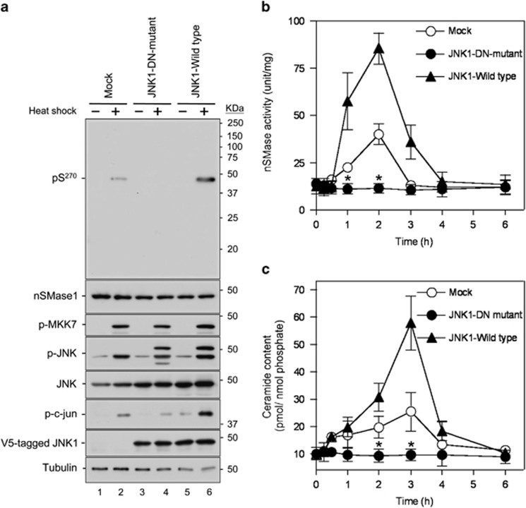
Figure 4.
Activation and phosphorylation of nSMase1 after the expression of a JNK DN mutant in heat-stressed zebrafish cells. (a) Detection of nSMase1 phosphorylation by the overexpression of JNK and its DN mutant (K55R). Mock cells (lanes 1 and 2), JNK1-DN (DN) mutant cells (lanes 3 and 4), and JNK1 wild-type cells (lanes 5 and 6) were either unstressed (normal growth, −) at 25 °C for 60 min, or heat-shocked (+) at 38 °C for 60 min. They were then analyzed by western blotting with antibodies against phosphorylated nSMase1, nSMase1, phosphorylated MKK7, phosphorylated JNK, JNK, phosphorylated c-jun, V5-tagged JNK1 wild-type, V5-tagged JNK1-DN mutant, and tubulin. Molecular weight markers are shown in kDa on the right. (b) Changes in nSMase activity. Mock, JNK1-DN, and JNK1 wild-type cells were heat-shocked at 38 °C for 0, 15, 30, or 60 min, allowed to recover at 25 °C for up to 5 h, and then harvested at the indicated times. nSMase activity was measured using C6-NBD-sphingomyelin as a substrate. Values represent the means of three independent experiments, and the error bars represent the S.D.s. *P<0.01 versus the mock control. (c) Changes in ceramide content. The ceramide content was measured using the diacylglycerol kinase assay. Values represent the means of three independent experiments, and error bars represent S.D.s. *P<0.01 versus the mock control

