Figure 3.
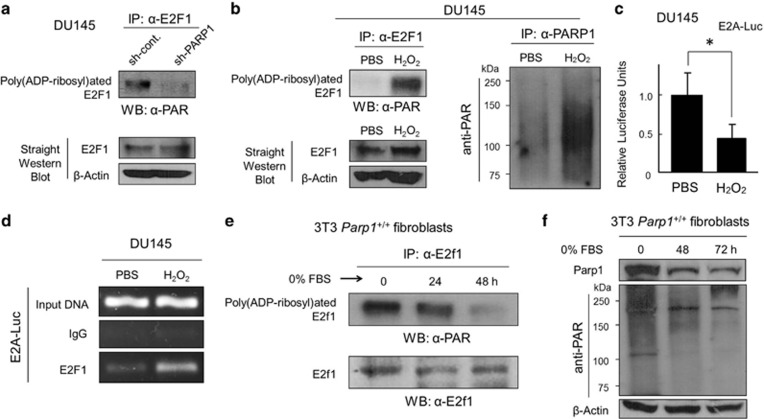
PARP1 modifies E2F1 by poly(ADP-ribosyl)ation. (a) IP/Western analysis of poly(ADP-ribosyl)ated E2F1 in DU145±sh-PARP1 cells. (b) IP/western analysis of poly(ADP-ribosyl)ated E2F1 in DU145 cells cultured with or without H2O2 (10 μM) for 30 min. (c) E2A–Luc assays in DU145 cells with or without H2O2 (10 μM) for 30 min. *P<0.05. (d) ChIP analysis of endogenous E2F1 protein on E2A–Luc promoter with or without H2O2 (10 μM) for 30 min. (e, f) The mouse 3T3 Parp1+/+ fibroblasts were incubated in culture medium containing 0% FBS for the times indicated. Pre-cleared protein lysates were subjected to either IP with an anti-E2f1 antibody followed by western analysis with an anti-PAR antibody or directly to western analysis with an anti-E2f1 antibody, an anti-Parp1 antibody, or an anti-PAR antibody

