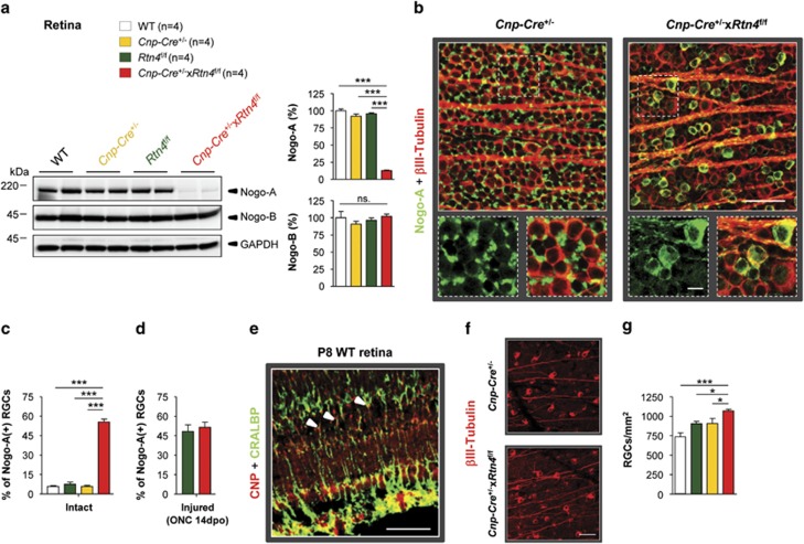
Figure 3.
Retinal ganglion cell body analysis reveals Nogo-A upregulation and higher cell survival in Cnp-Cre+/−xRtn4flox/flox KOs. (a) In the retina, Nogo-A expression was downregulated by ∼85%, whereas Nogo-B was unchanged in the Cnp-Cre+/−xRtn4flox/flox KOs. (b) In intact, Cnp-Cre+/− control retinae, Nogo-A was mostly located in Müller cell end-feet surrounding βIII-Tubulin-labelled retinal ganglion cell bodies. In contrast, in Cnp-Cre+/−xRtn4flox/flox KOs, Nogo-A expression was abolished in Müller cells and was strongly upregulated in RGCs. (c) Quantitatively, ∼7% of RGCs expressed Nogo-A in intact control retinae, whereas ∼55% of RGCs expressed the Nogo-A protein in the glial Nogo-A KOs (n=4 per group). (d) Two weeks after ONC injury the density of RGCs expressing Nogo-A was not significantly different between Rtn4flox/flox and Cnp-Cre+/−xRtn4flox/flox mice (n=3 per group). (e) In the retina, the CNP protein was expressed in Müller cells identified by using the cell-type specific marker CRALBP. (f) Two weeks after injury, the density of surviving RGCs was evaluated by immunostaining for βIII-Tubulin on retinal flat-mounts. (g) Retinal ganglion cell survival was slightly, but significantly increased in Cnp-Cre+/−xRtn4flox/flox KO animals compared with the control genotypes (n=3–7). Statistics: one-way ANOVA, Bonferroni's multiple comparison test, *P<0.05, **P<0.01, ***P<0.001. Scale bars: (b, e, f)=50 μm; (b) insets=10 μm

