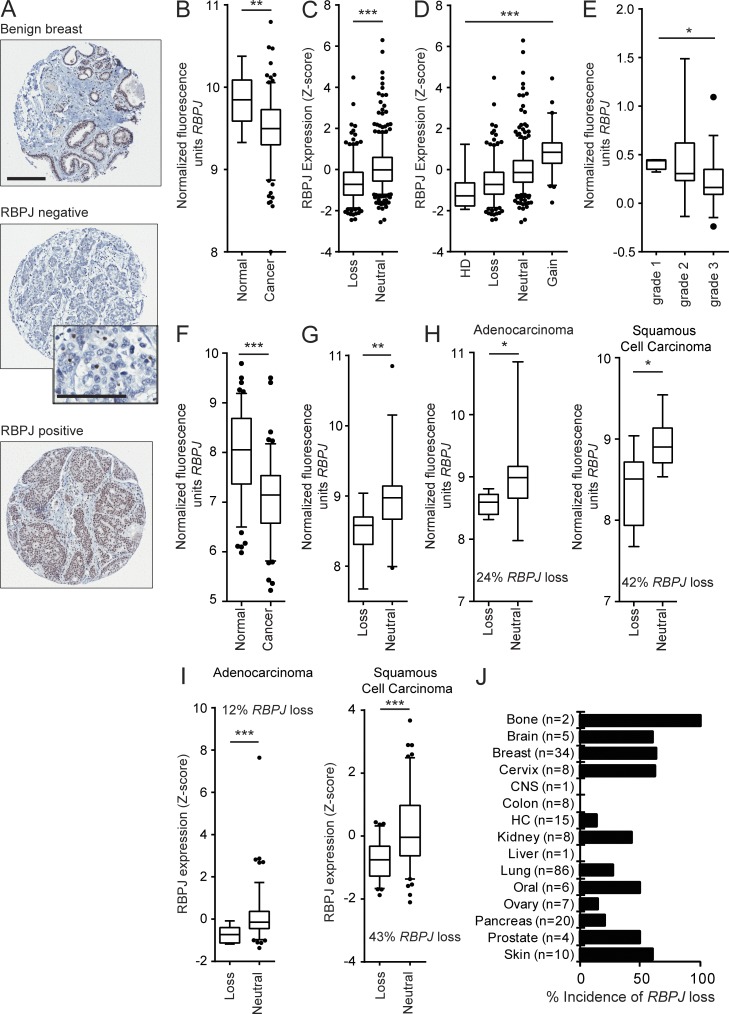
Figure 1.
RBPJ is frequently lost in human cancers. (A) Examples of RBPJ immunohistochemical staining in benign breast tissue (n = 8) and breast cancer tissue microarray cores (RBPJ negative, n = 40; RBPJ positive, n = 224; bar, 200 µm). High power inset (bar = 100 µm) of the RBPJ-negative tumor core shows positive staining in internal control cells in the tumor microenvironment. (B) RBPJ mRNA expression in breast tumors (n = 183) and adjacent normal breast tissue (n = 13; Yu et al., 2008). (C) Analysis of RBPJ expression and RBPJ genomic copy loss (n = 277) versus no loss (neutral, n = 551) in invasive breast cancers (TCGA data). (D) Data from C plotted by RBPJ copy number status; HD (n = 7), loss (n = 270), neutral (n = 489), and amplification (gain, n = 62). P < 0.0001 by Kruskal–Wallis followed by Dunn’s multiple comparisons post-test showed significant differences in all comparisons except between the HD versus loss group. (E) RBPJ mRNA expression in human breast cancers stratified by tumor grade; grade 1 (n = 4), grade 2 (n = 12), and grade 3 (n = 39; Ginestier et al., 2006). (F) RBPJ mRNA expression in normal bronchial epithelium collected from healthy individuals (n = 67) versus non–small cell lung carcinoma (n = 111; Bild et al., 2006; Lockwood et al., 2010). (G) Analysis of lung cancers of mixed type with RBPJ genomic copy loss (n = 14) versus no loss (n = 30) with paired mRNA expression and aCGH data (Lockwood et al., 2008, 2010). (H) Analysis of lung cancers from G broken down into tumor subtypes; adenocarcinoma RBPJ genomic loss (n = 6) versus no loss (n = 19); squamous cell carcinoma RBPJ genomic loss (n = 8) versus no loss (n = 11; Lockwood et al., 2008, 2010). (I) Analysis of RBPJ mRNA expression and genomic copy number in TCGA lung cancer adenocarcinomas (RBPJ genomic loss [n = 15] versus no loss [n = 114]) and squamous cell carcinomas (RBPJ genomic loss [n = 77] versus no loss [n = 101]; Cerami et al., 2012; Lockwood et al., 2012; Gao et al., 2013). (J) RBPJ copy number alteration evaluated using aCGH across a panel of 215 cancer cell lines (CNS: central nervous system, HC: hematopoietic cell lines). At least one allele of RBPJ is lost at an overall frequency of 35% (also see Table S1). P-values were obtained using a Mann-Whitney test for B, C, and F–I and a Kruskal-Wallis one-way analysis of variance test for D and E. Data are presented as box-and-whisker plots. The bottom and top bars of the whisker indicate the 5th and 95th percentiles respectively with points outside the whiskers shown as individual dots. *, P < 0.05, **, P < 0.005, ***, P < 0.0001.

