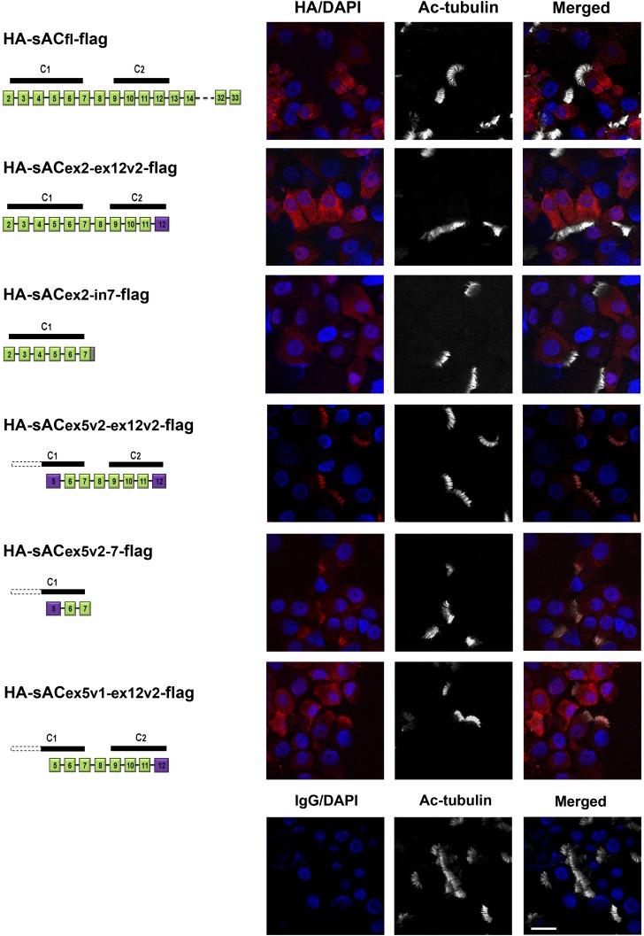
Figure 3.
Localization of sAC isoforms in NHBE cells. Lentivirus constructs expressing different portions of sAC with an N-terminal HA and C-terminal flag tag were used to infect undifferentiated NHBE cells. After the cells were differentiated using ALI conditions, the location of the expressed sAC was determined in cytospin preparations using HA antibodies and immunofluorescence, shown to the right of each construct. These are representative images of each construct tested in cells from at least three donors. Left: diagrams of sAC constructs expressed in NHBE cells. Green boxes represent the coding exons, purple boxes indicate newly classified exons (containing previously thought intron sequences), and the gray box shows an added intron stop codon. White dashed bars indicate the missing catalytic domain, and black bars represent the contained catalytic domains. Right panels: Immunofluorescence of cytospin preparations of fully differentiated NHBE cells infected with lentiviruses expressing different sAC variants, stained with mouse anti-HA antibody (red), cilia with rabbit anti–acetylated tubulin (Ac-tubulin) antibody (white), and 4′,6-diamidino-2-phenylindole (DAPI) for nuclei (blue). HA-sACfl-flag, HA-sACex2-ex12v2-flag, and HA-sACex2-in7-flag are localized in the cytoplasm of ciliated cells. HA-sACex5v2-ex12v2-flag and HA-sACex5v2-in7-flag are localized to cilia. HA-sACex5-ex12v2-flag is found in both cilia and cytoplasm. Lower right panels: NHBE cells infected with the lentivirus vector with no insert served as a negative staining control. Scale bar, 20 μm.

