Figure 4.
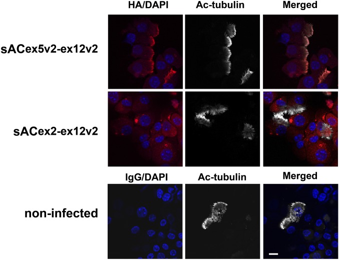
Localization of sAC variants in C2 knockout (KO) murine airway epithelial cells. HA-sACex5v2-ex12v2-flag or HA-sACex2-ex12v2-flag were infected into undifferentiated sAC C2 KO murine airway epithelial cells. Cytospin preparations were made from fully differentiated airway epithelial cells and stained with an HA antibody (red). Cilia were identified with an acetylated tubulin (Ac-tubulin) antibody (white). DAPI shows nuclei (blue). These are representative of two or more independent experiments. Upper panels: the HA-sACex5v2-ex12v2-flag variant is localized to cilia, analogous to NHBE cells. Middle panels: the HA-sACex2-ex12v2-flag variant is not localized to cilia, but remains in the cytoplasm, again analogous to NHBE cells. Lower panels: noninfected C2 KO murine cells stained with mouse IgG antibody. Scale bars, 10 μm.

