Figure 5.
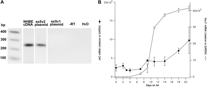
Expression of ex5v2 containing sAC variants during differentiation. (A) Agarose gel analysis of PCR products using ex5v2-specific primers (see Table 1, last pair). Lane 2, cDNA from fully differentiated NHBE cells; lane 3, a plasmid with ex5v2 variant; lane 4, a plasmid with ex5v1 variant (i.e., without the retained intron 4 sequence); lane 5, with no reverse transcriptase; lane 6, with H2O only. The expected 266-bp band is observed in the human cell cDNA and ex5v2 plasmid, but not in the ex5v1 plasmid, indicating that the primers are specific for the ex5v2 splice variant. DNA size markers are in lane 1. (B) Graph of the ex5v2 expression (closed circles) during NHBE cell differentiation relative to glyceraldehyde 3-phosphate dehydrogenase (GAPDH) mRNA measured by SYBR green quantitative RT-PCR. The level of FoxJ1 mRNA (open circles) is shown as a marker for ciliated cell differentiation. The amount of ex5v2 mRNA increases roughly threefold during NHBE cell differentiation. Data are shown as mean (± SEM).

