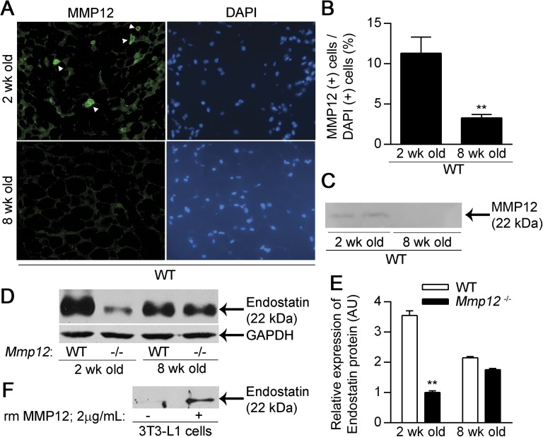
Figure 3.
MMP12 and endostatin are increased in the white adipose tissue of 2–week-old mice. WT mice were killed at 2 and 8 weeks of age, and the subcutaneous adipose tissue was analyzed for MMP12 expression (n = 4 mice/group). (A) Representative staining for MMP12. (B) The number of MMP12-expressing cells as a percentage of DAPI+ cells. (C) Representative Western blot for MMP12 protein expression in 60 μg of protein from WT and Mmp12−/− subcutaneous adipose. Endostatin levels were analyzed in the subcutaneous adipose tissue of 2- and 8-week-old WT and Mmp12−/− mice (n = 3 mice/group). (D) Representative Western blot for endostatin protein and glyceraldehyde 3-phosphate dehydrogenase (GAPDH) endogenous control. (E) Endostatin levels were quantified by densitometry. (F) Representative Western blot demonstrating cleavage of Collagen XVIII to endostatin in 3T3-L1 adipocytes treated with recombinant murine (rm) MMP12. Data are mean ± SEM. **P < 0.005 versus 8-week-old mice from respective strain.

