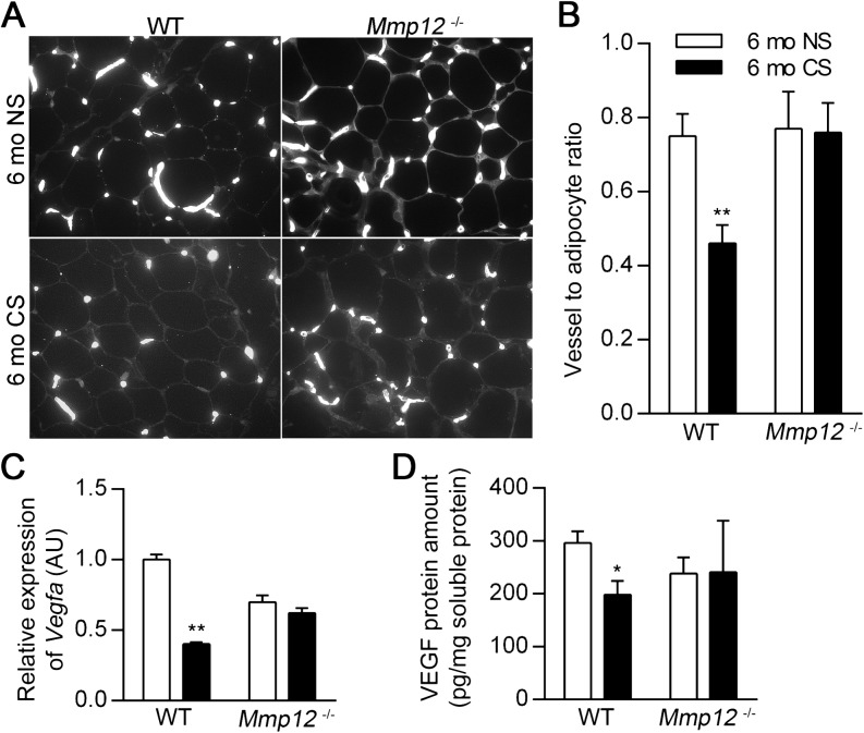
Figure 6.
WT mice have decreased adipose vasculature after 6 months of CS exposure. Subcutaneous adipose tissue was harvested from WT and Mmp12−/− mice after 6 months of exposure to NS or CS. Representative blood vessel histology by lectin staining (A) and vessel-to-adipocyte ratios in subcutaneous adipose tissue (B) (n = 5 mice/group). Vegfa mRNA (C) and VEGF protein (D) were quantified by real-time RT-PCR and ELISA, respectively, in subcutaneous adipose tissue (n = 4–7 mice/group). VEGF ELISA values were normalized to total soluble protein concentration. Data are mean ± SEM. *P < 0.05 and **P < 0.005 versus respective NS controls.

