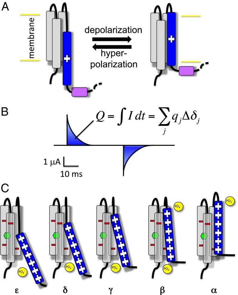
Fig. 1.
Activation of voltage sensor domains (A) VSDs are formed by four transmembrane helices. Upon changes in the membrane potential, the positively charged S4 (blue) moves across the membrane relative to a static S1–S3 bundle (gray), transmitting the electrical signal to a linker peptide (purple). (B) This movement is reported by the measurement of transient currents called gating currents. The time integral of these, the gating charge , can be expressed as the sum of the contributions of the charges of the system. (C) Cartoon depiction of the stepwise activation of the Kv1.2 VSD. From the most resting (ε) to the most activated conformation (α), S4 proceeds in a ratchet-like upward motion in which its positively charged residues jump from a negative binding site to the next. The negative charges of S1–S3 are depicted in red and the ones of the lipid headgroups in yellow. The hydrophobic gasket at the center of the VSD is represented by a green hexagon.

