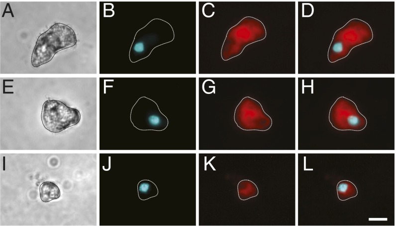
Fig. 2.
Morphology of spinning cells. Dissociated cells were stained with the membrane marker FM1-43 and the nuclear Hoechst stain to confirm the cellular nature of spinning objects. (A, E, and I) Representative transmitted light images of magnetic cells from the lagena (A and E) and the olfactory epithelium (I). (B, F, and J) Fluorescent images of cells stained with Hoechst (shown in cyan). (C, G, and K) Fluorescent images of cells stained with FM1-43 (shown in red). (D, H, and L) Shows merged images. The periphery of cells (dashed lines) was defined by the FM1-43 staining. (Scale bar: in L, 10 μm.)

