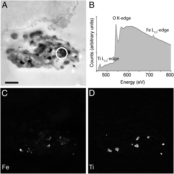
Fig. 4.
Elemental analysis of spinning cells. (A) High-magnification TEM micrograph of the electron-dense extracellular structure associated with the spinning cell shown in Fig. 3 E and F. The structure shown in Fig. 4A is a mirror image of that shown in Fig. 3 E and F, as the image was captured on a different microscope. (Scale bar: 1 μm.) (B) Background-corrected electron energy loss spectra (EELS) obtained from the circled region in A. Titanium, oxygen, and iron were detected. Using energy-filtered transmission electron microscopy (EFTEM), element maps were generated for iron (C) and titanium (D). These elements are found throughout the extracellular aggregate, strongly suggesting that they are contaminants.

