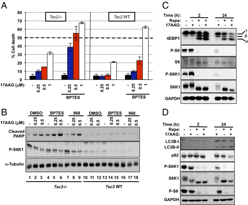
Fig. 3.
Inhibition of glutamine anaplerosis and Hsp90 causes potent apoptosis in Tsc2−/− cells. (A) Cell death of Tsc2−/− (Left) and Tsc2–WT (Right) MEFs after 72 h of treatment with increasing concentrations of 17AAG with or without BPTES (10 μM) was measured via propidium iodide (PI) exclusion assay. The mean is shown; error bars represent SEM (n > 3). (B) Immunoblot analysis of cleaved PARP, P-S6K1, 4E-BP1, and α-tubulin in Tsc2−/− MEFs treated with the indicated compounds for 24 h. (C) Immunoblot analysis of downstream targets of the mTORC1 pathway in Tsc2−/− MEFs treated with rapamycin (20 ng/mL), 17AAG (1 μM), or the combination of both for the indicated time points. (D) Immunoblot analysis of LC3B, p62, P-S6K1, S6K1, P-S6, and GAPDH in Tsc2−/− MEFs treated as in C.

