Figure 5.
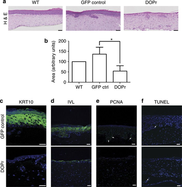
δ-Opioid receptor (DOPr) activity results in decreased epidermal thickness and atypical stratification. Organotypic cultures of N/TERT-1 cells non-transduced or transduced with control or DOPr-green fluorescent protein (GFP) lentivirus were generated. (a) Hematoxylin and eosin (H&E) staining sections of organotypic raft cultures show decreased epidermal thickness when the DOPr was overexpressed. Bar = 100 μm. (b) Epidermal areas from H&E staining were quantified and normalized to the respective wild-type (WT) control. Graph represents the mean±SD (n=3) with duplicates per experimental condition. One-way analysis of variance, *P<0.05. Tissue sections were labeled for differentiation markers (c) keratin intermediate filament (KRT10) and (d) involucrin (IVL) by immunofluorescence. (e) Proliferating cell nuclear antigen (PCNA) immunofluorescence labeling marks proliferating cells in the basal layer of control, and (f) the TUNEL assay shows only single apoptotic cells in both control and DOPr-overexpressing cultures. This correlates well with the atrophic phenotype. Nuclei were counterstained with Hoechst. Bar=50 μm.

