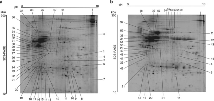
Figure 1.
Images of representative 2D gels. The images are representative of six samples showing separation of proteins extracted from lesions of (a) cutaneous leishmaniasis (CL) patients and (b) normal skin. Directions of isoelectric focusing and SDS-PAGE are indicated on the top and on the side of the left gel image. Spots marked only in gel (a) indicate protein spots unique in the CL samples. Spots marked only in gel (b) show the protein spots unique in normal skin. Spots marked in gels (a, b) indicate protein spots differently regulated between the samples. The protein spots were identified by matrix-assisted laser desorption/ionization time of flight (MALDI-TOF) mass spectrometry. List of identified proteins is given in Table 1.

