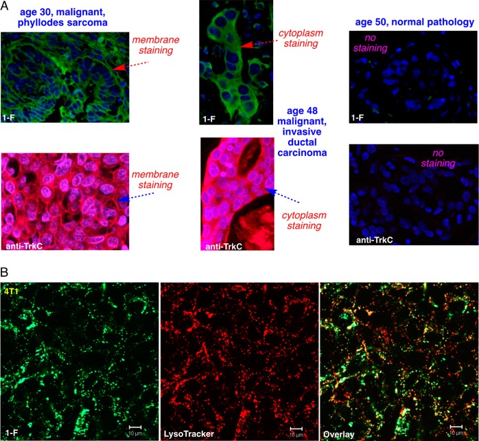
Figure 2.
Compound 1-F stains in TrkC+ tumor tissue and is internalized TrkC+ cells. (A) Histochemical stains for a library of 96 breast tissue slices were performed using 1-F (top) and anti-TrkC antibody as control (bottom), and the three illustrative ones shown here illustrate staining of the malignant tumor, whereas normal tissue is not stained. No staining was observed in the tissues without the small molecule probe or mAb. (B) Cell imaging on 4T1 cells shows 1-F was internalized into lysosomes just as the natural TrkC ligand NT3 is.

