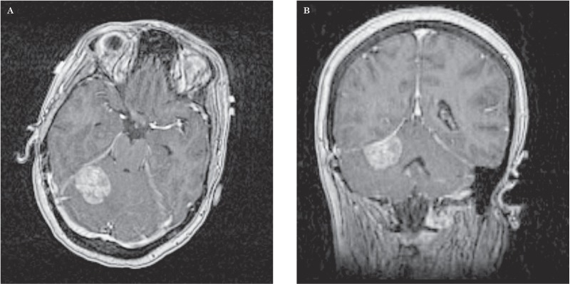
Figure 2.
Axial (A) and coronal (B) contrast-enhanced T1-weighted MRI of the brain revealed a 2.5 × 3.0 × 1.7 cm dural-based, extra-axial, enhancing soft tissue mass arising from the right tentorium cerebelli with vasogenic edema and mild mass effect upon the right cerebellar hemisphere and fourth ventricle. The lesion was favored to represent a meningioma. Given the patient's history of cancer, however, a solitary broad-based dural metastasis could not be excluded.

