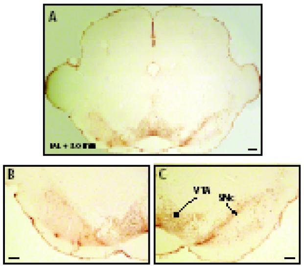
Fig. 2.
Representative TH-immunostained left side SNc MPTP lesion. Panel A shows the lesion on the left side and sham on the right side. Panel B shows detail of the VTA and SNc of lesioned side, panel C the SNc and VTA of the sham side. SNc, substantia nigra compacta; VTA, ventral tegmental area. Scale bar represents 200 μm.

