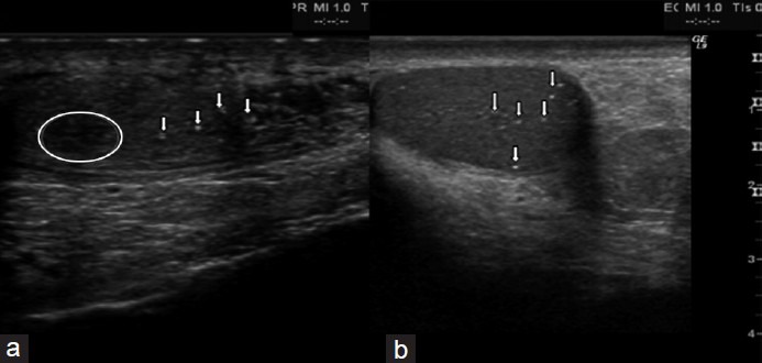
Figure 3.

Longitudinal scrotal ultrasonography of two Klinefelter syndrome subjects (a) widespread inhomogeneous testicular structure with micro-calcifications (arrows) and small hypo-echoic areola (circled). (b) Various testicular micro-calcifications (arrows) in an inhomogeneous testicular echo-structure.
