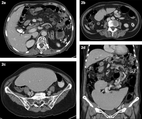
Fig. 2.

(a–c) Contrast-enhanced axial CT images of the abdomen and pelvis demonstrate the ‘whorl’ sign of the long vascular pedicle extending to the abnormally positioned spleen. There is no fat stranding surrounding the twisted splenic pedicle, and contrast enhancement of the splenic parenchyma is preserved with no evidence of infarction. Gallstones are also noted. (d) Contrast-enhanced coronal reconstructed CT image demonstrates a lobulated, enlarged spleen, which lies in the pelvis and lower abdomen, with an elongated splenic pedicle in torsion.
