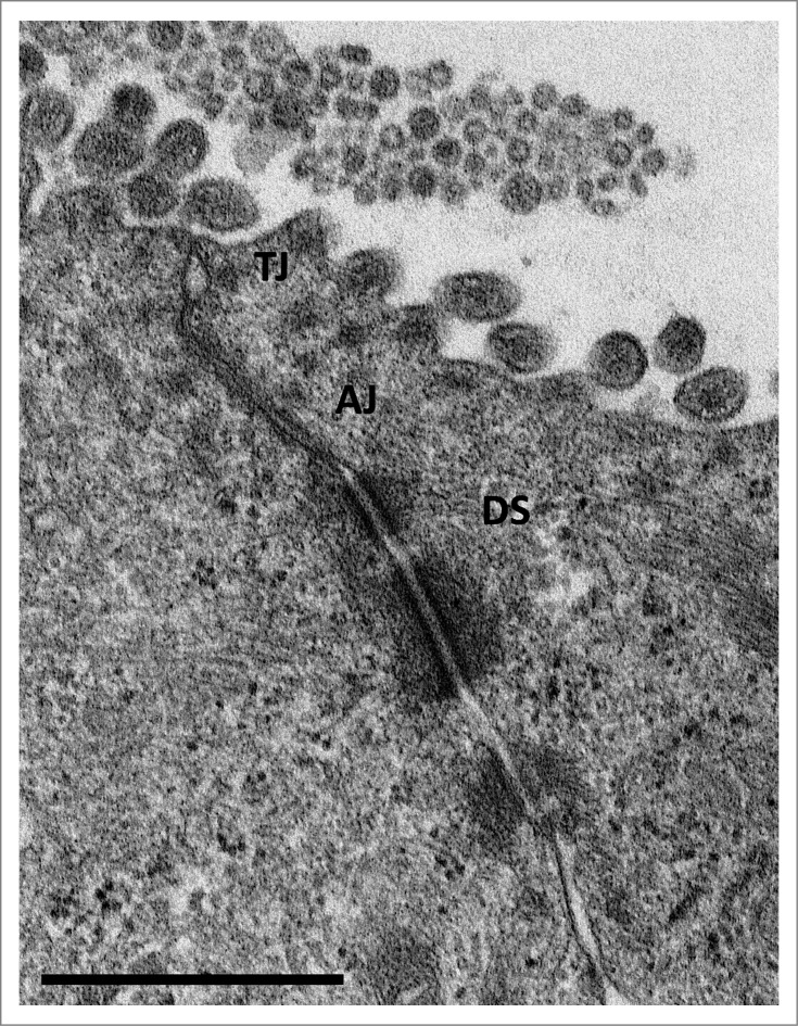
Figure 1.
Transmission electron microscopy (TEM) image of an 'apical junctional complex' of polarized T84 human colorectal carcinoma epithelial cells Depicted are the ‘tight junctions’ (TJ), directly beneath the microvilli, ‘adherens junctions’ (AJ), and ‘desmosomes’ (DS) below the ‘apical junctional complex’. Scale bar = 1 μm (courtesy of Lilo Greune, Institute of Infectiology – Center for Molecular Biology of Inflammation (ZMBE), University of Münster).

