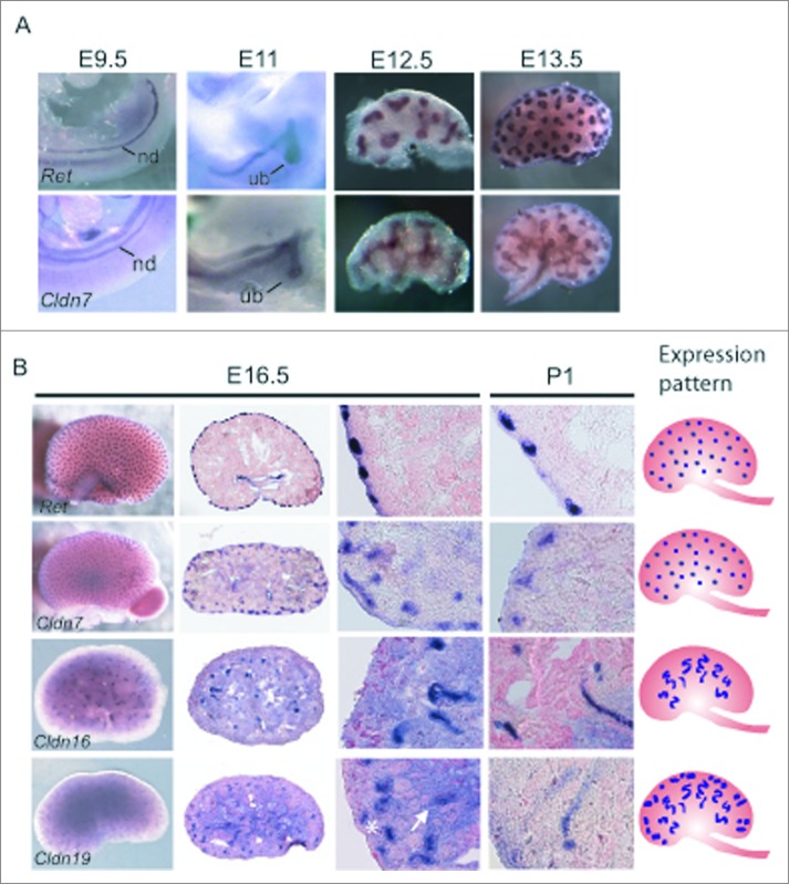
Figure 2.
Spatial Expression Analysis of Cldn7, Cldn16, and Cldn19 mRNA Transcripts during Mouse Kidney Development. (A-B) Whole mount in situ hybridization was performed on whole embryos at embryonic day (E) 9.5 and E11.0, and on whole kidneys at E12.5, E13.5, E16.5, and P1 for Cldn7, Cldn16, Cldn19, and Ret. E16.5 and P1 kidneys were cryosectioned at 15 μm thickness and counterstained with eosin. Cldn7 expression is observed in the nephric duct (nd) at E9.5 and in the ureteric bud (ub) at E11.0. At E11.0, E12.5, E13.5 and E16.5 Cldn7 is seen in the ureteric bud trunks and the tips. At P1, Cldn7 is detected mostly in ureteric bud tips, similar to Ret expression at the same stage. Cldn16 and Cldn19 transcripts are first detected at E16.5. Cldn16 is predominantly expressed in late tubular structures, while Cldn19 is expressed in both early and late tubular structures that will eventually become the Loop of Henle. On the far right schematic patterns are shown indicating a ureteric bud tip pattern (Cldn7 and Ret), a late tubule pattern (Cldn16), and a combined early and late tubule pattern (Cldn19) as described.23

