Figure 1.
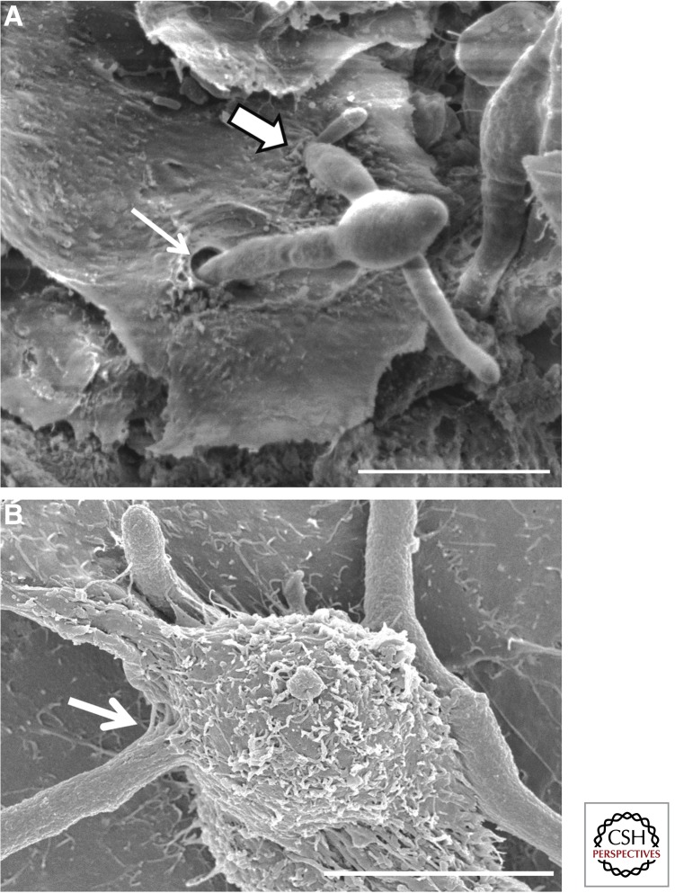
Invasion of epithelial cells by C. albicans and Aspergillus fumigatus. Scanning electron micrograph of (A) C. albicans invasion of the epithelial cells on the dorsum of the tongue of a cortisone acetate–treated mouse with oropharyngeal candidiasis. Thick arrow indicates a hypha that is likely invading by induced endocytosis, which is characterized by ruffling of the epithelial cell plasma membrane at the site of invasion. Thin arrow indicates a hypha that is probably invading by active penetration. Note the absence of membrane ruffling at the invasion site. (Image courtesy of Dr. Mary Ann Jabra-Rizk, University of Maryland.) (B) A. fumigatus invasion of A549 pulmonary epithelial cells. Arrow indicates a hypha penetrating the epithelial cell surface, likely by induced endocytosis. Scale bars, 10 µm.

