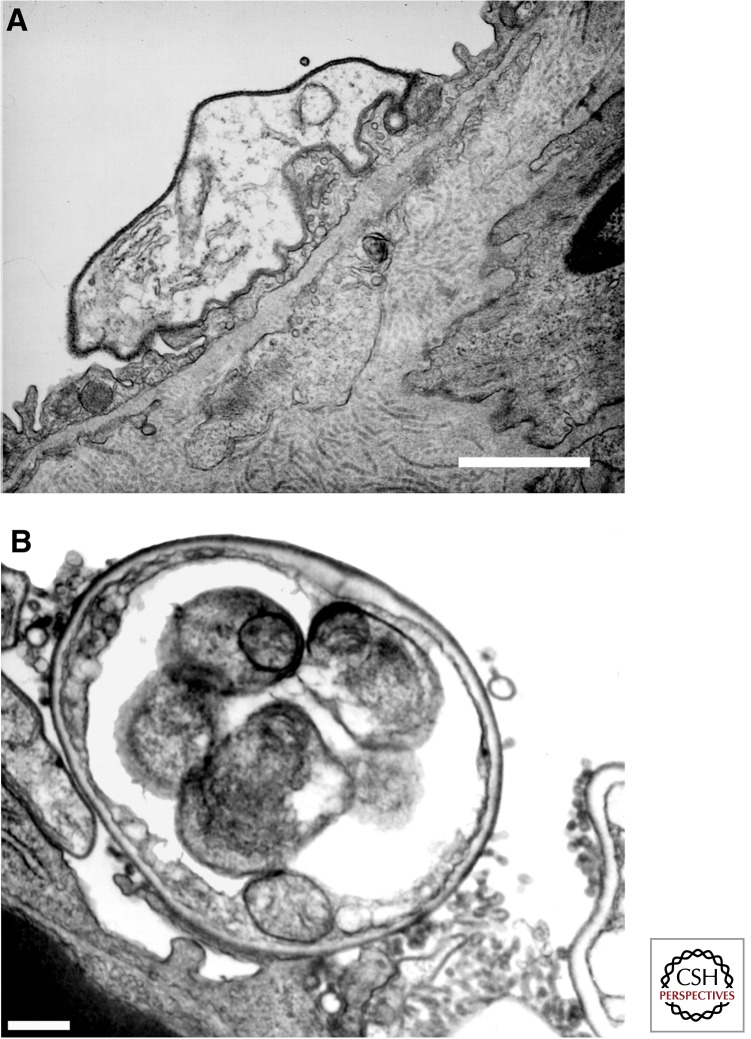
Figure 1.
Electron micrographic visualization of typical Pneumocystis life cycle forms. (A) Reveals a trophic form of P. carinii adherent to a type I alveolar epithelial cell, mediated by interdigitation of the organism’s plasma membrane with that of the host cell. (B) Shows a thick-walled cyst form of P. carinii. Scale bars, ∼1 μm. (From Limper et al. 1997; reprinted, with permission, from the author.)

