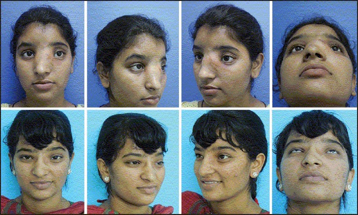
Figure 14.

A case of frontonasal meningo-encephaloceles showing telecanthus (upper row). The correction was done using Chula technique and the medial orbital wall was reconstructed along with medial canthopexy. The postoperative appearance 3 years later is shown in the lower row
