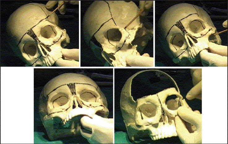
Figure 6.

Markings for the orbital box osteotomy on a skull model. The frontal craniotomy and the various markings for the cuts in the orbital walls and roof are shown in upper row and the left picture in lower row. The picture on right in the lower row shows the moved orbits after removal of central excess tissue and completion of box osteotomy cuts (pictures courtesy Dr. Mukund Jagannathan)
