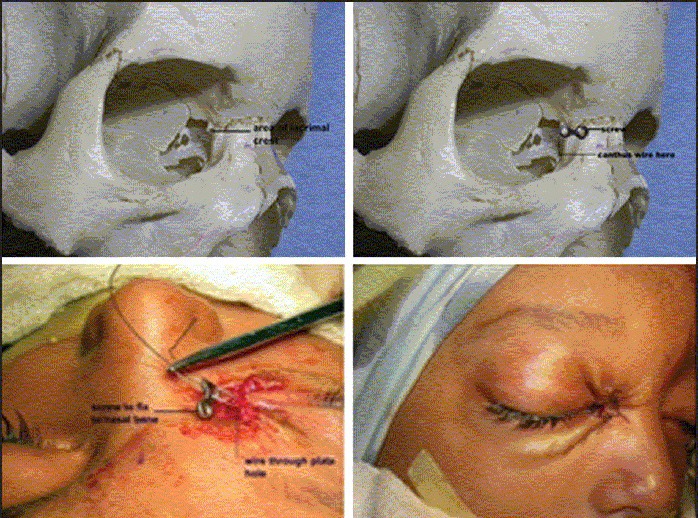
Figure 8.

Author's technique of medial canthopexy. The upper row (left) shows the position of lacrimal crest in a skull. The picture on right shows placement of a two-hole plate with the lower hole at the level of posterior lacrimal crest. The lower row (left shows threading of the canthal wire into the lower hole prior to fixing the plate at the upper hole with a screw to the solid nasal bone
