Figure 4.

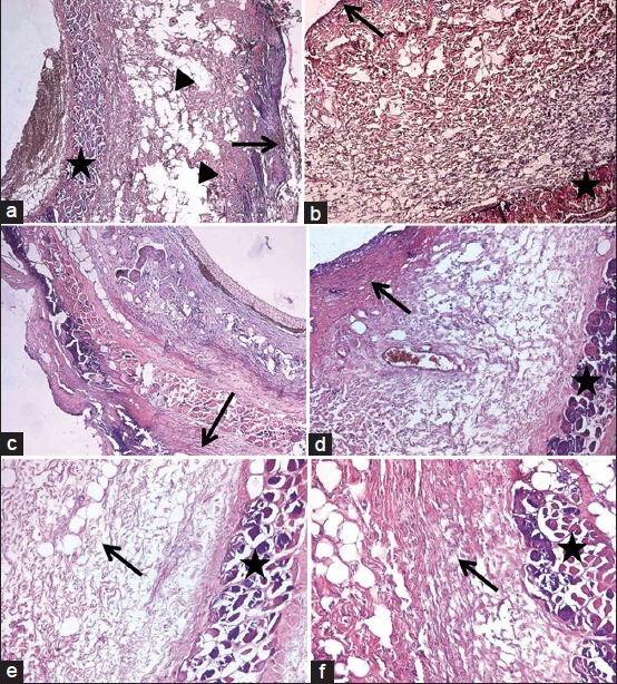
(a) Microscopic pictures of the groups: Negative pressure wound therapy (NPWT) group at 3rd day; a thin layer of granulation tissue (arrow) above edematous areas (arrow head) between the thick collagen fibers and deeply located necrotic muscle fibers (star). (b) Microscopic pictures of the groups: Dressing group at 3rd day; granulation tissue is similar to NPWT group but with thin collagen fibers underneath (arrow: Granulation tissue, star: Necrotic muscle fibers). (c) Microscopic pictures of the groups: Dressing group at 3rd day; granulation tissue is similar to NPWT group but with thin collagen fibers underneath (arrow: Granulation tissue, star: Necrotic muscle fibers). (d) Microscopic pictures of the groups: Dressing group at 7th day; thick granulation tissue composed of irregularly arranged thin collagen fibers (arrow) above the necrotic muscle fibers (star). (e) Microscopic pictures of the groups: NPWT group at 14th day; edematous connective tissue composed of collagen fibers resembling dermis (arrow) and necrotic muscle fibers (star). (f) Microscopic pictures of the groups: Dressing group at 14th day; collagen fibers oriented as scar tissue (arrow) above necrotic muscle fibers (star)
