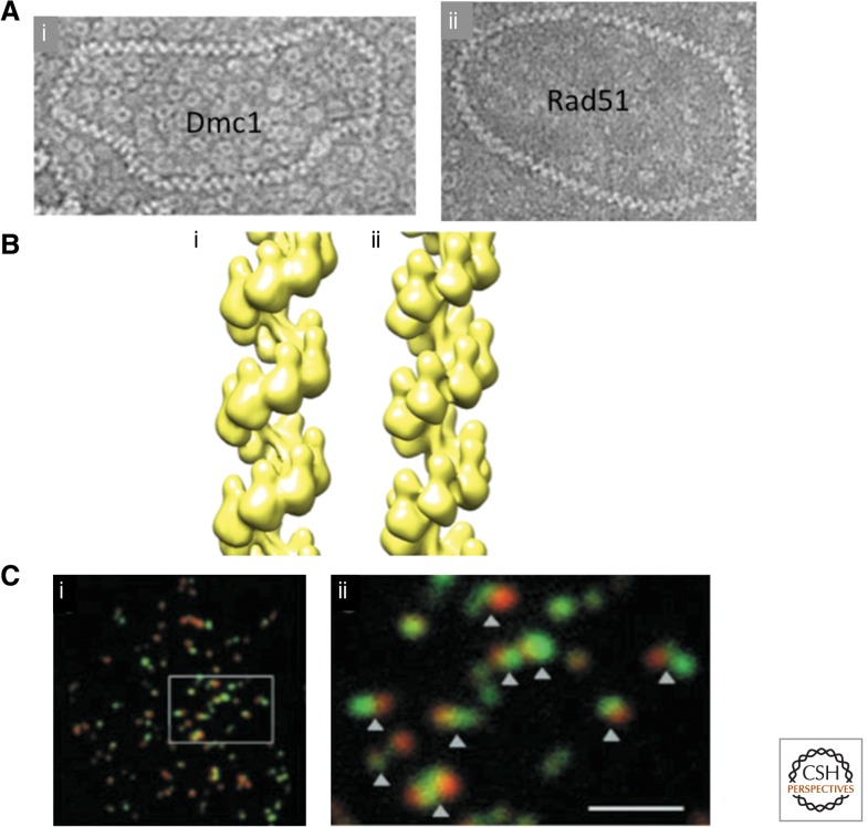
Figure 2.
Microscopic analysis of strand-exchange proteins. (A) Electron micrographs of human (i) Dmc1, and (ii) Rad51filaments coating a 1312 bp circular dsDNA plasmid. Note the high density of toroids in the background of the Dmc1 image (From Sheridan et al. 2008; reprinted, with permission, from Oxford University Press © 2008.) (B) Helical reconstructions of human (i) Dmc1, and (ii) Rad51 filaments (courtesy of E. Egelman). (C) Surface spread S. cerevisiae meiotic nuclei immunostained for Rad51 (green) and Dmc1 (red). (i) Low magnification view, and (ii) blow up of region indicated. Scale bar, 2 μm. (From Shinohara et al. 2000; reprinted, with permission, from the American Society of Plant Biologists © 1999.)

