Fig. 3.

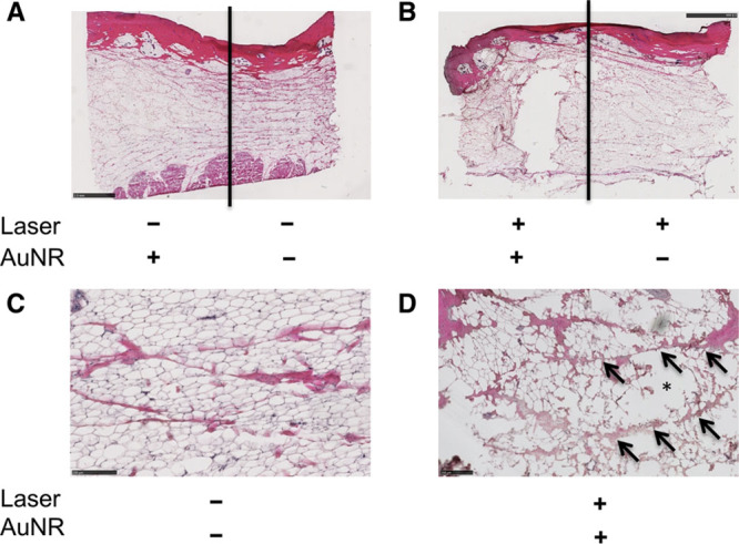
Histological effects of NanoLipo in ex vivo porcine skin and subcutaneous tissue. A, Hematoxylin and eosin (H&E)-stained section of AuNR-injected (left) and untreated (right) samples. B, H&E-stained section of NanoLipo-treated (left) and laser-treated samples. Higher magnification power image of untreated region (C) and NanoLipo-treated region (D); arrows indicate intact connective tissue and asterisk indicates disruptions. Scale bars = 2.5 mm (A and B), 250 μm (C and D).
