Figure 5.

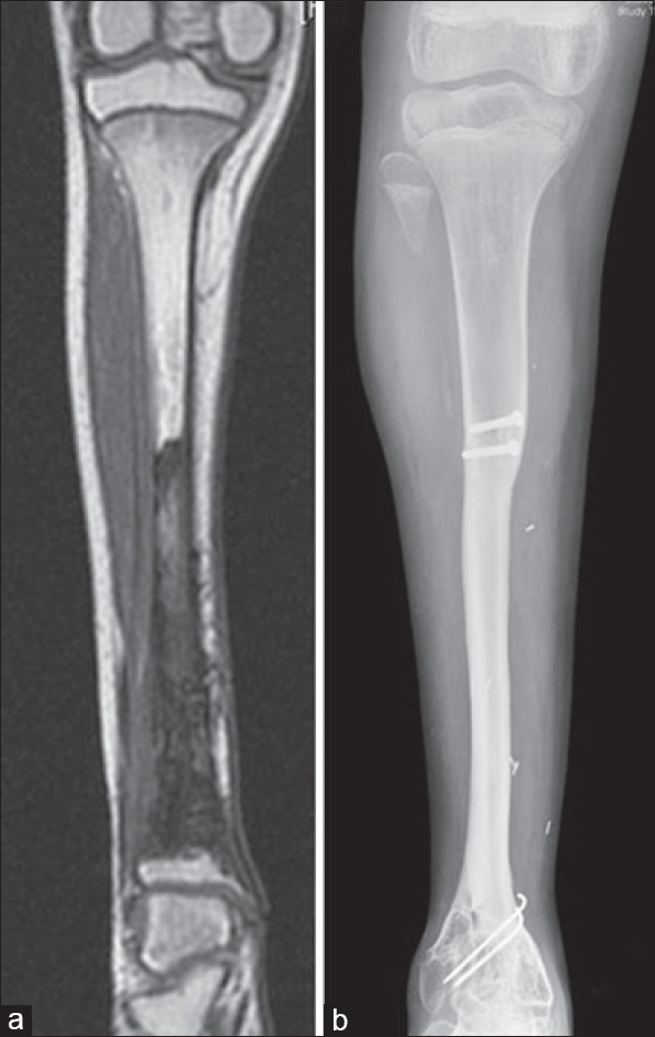
(a) Preoperative magnetic resonance imaging of tibia showing an Ewing sarcoma (b) followup radiograph at 36 months showing hypertrophy of the transposed (medial translation into post excision defect) fibula

(a) Preoperative magnetic resonance imaging of tibia showing an Ewing sarcoma (b) followup radiograph at 36 months showing hypertrophy of the transposed (medial translation into post excision defect) fibula