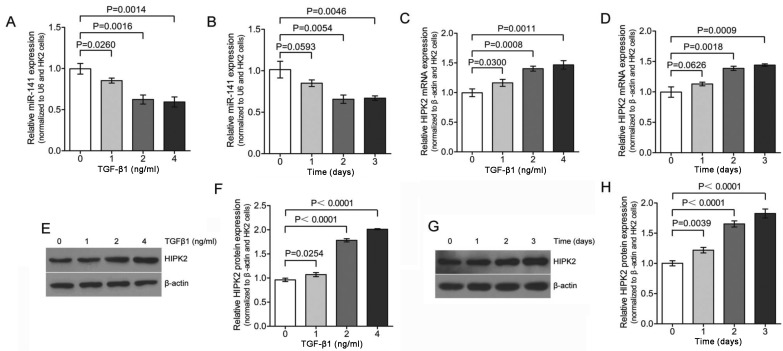
Figure 2.
TGF-β1 induces downregulation of miR-141 accompanied by up regulation of HIPK2. HK-2 cells were treated with (A) TGF-β1 (0, 1, 2 and 4 ng/ml for 2 days) or (B) TGF-β1 (2 ng/ml) for 0–3 days. The expression of miR-141 was assessed by qPCR and normalized to U6 expression. The expression for the various dose points was determined as a relative change in the absence of TGF-β1 treatment and shown as mean ± standard error (S.E). HK-2 cells were incubated with (C and E) TGF-β1 (0, 1, 2 and 4 ng/ml for 2 days) or (D and G) TGF-β1 (2 ng/ml) for 0–3 days and the HIPK2 expression level was assessed by (C and D) qPCR and (E and G) western blotting, which were consistent with the RNA expression analysis in C and D, respectively. (F and H) The results from the western analysis in E and G were quantified and subjected to densitometry and shown as a graph (P-values are compared to the control). The relative expression of HIPK2 was normalized to β-actin expression and the expression for the various dose points was determined as a relative change from dose 0 and shown as mean ± S.E. TGF, transforming growth factor; HIPK2, homeodomain-interacting protein kinase 2; qPCR, quantitative polymerase chain reaction.

