Figure 5.
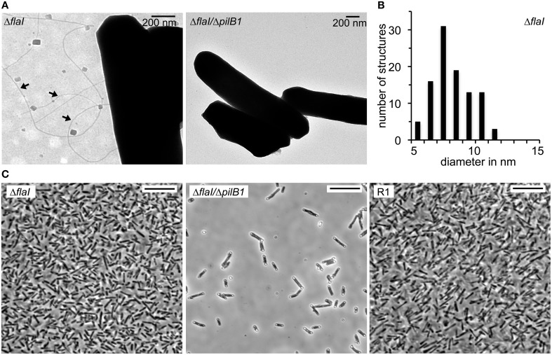
Phenotypic characterization of the Hbt. salinarum R1, ΔflaI and ΔflaI/ΔpilB1 mutant strains. (A) Transmission electron micrographs of surface attached cells on carbon coated gold grids after 10 days of cultivation at 42°C. Pili-like surface structures observed with ΔflaI are labeled with arrows. (B) Frequency distribution of 100 filaments diameters in nm found with the ΔflaI mutant. (C) Light micrographs of Hbt. salinarum R1 and mutant strains attached to a glass surface after 10 days of cultivation at 42°C. Cells not attached to the surface were removed by stringent washing. Scale bars are 10 μm.

