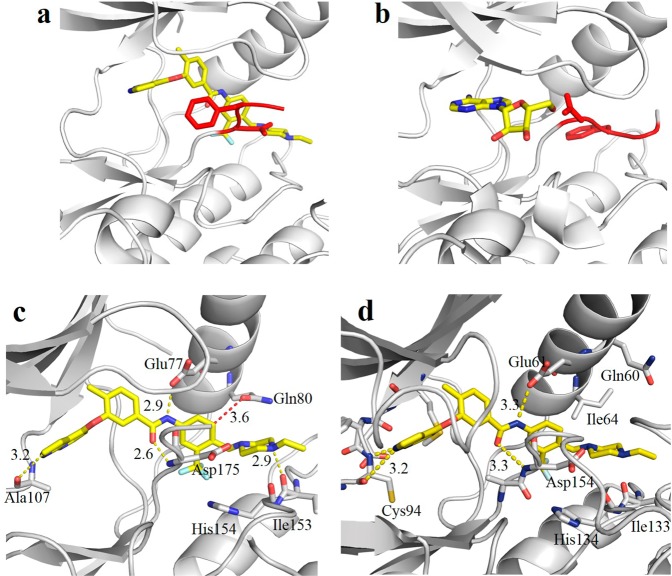
Figure 3.
Compound 1 is a type II inhibitor. (a) Binding of 1 to the active site of TAK1–TAB1 results in the DFG-out conformation characterized by type II inhibitors. (b) The structure of adenosine bound to the active site of TAK1–TAB1 (PDBID 2EVA) is provided for comparison and shows the DFG-in conformation. The DFG motif is highlighted in red. (c) Key interactions of 1 with TAK1. (d) Molecular model of the binding mode of MAP4K2 with 1.

