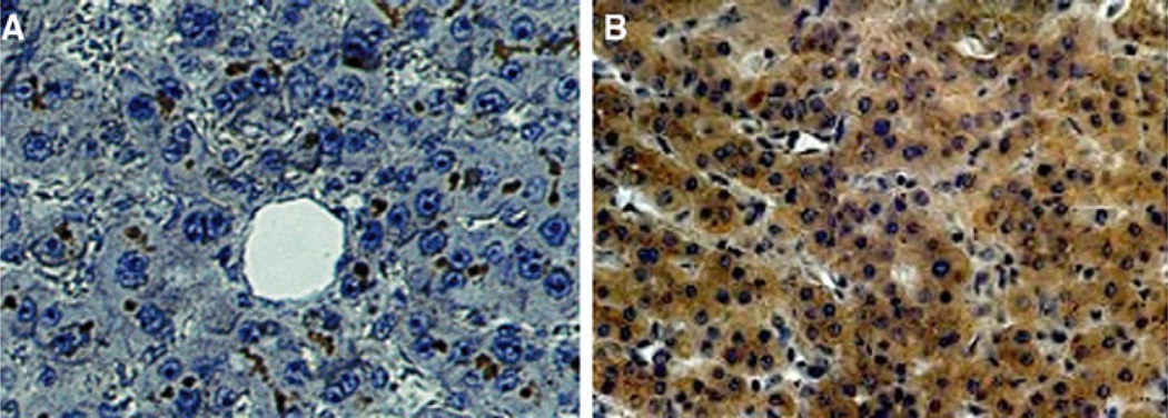
Fig. 5.
Distribution of CEACAM1 in human liver. There is no CEACAM1 expression in cotton top tamarin (CTT) and common marmoset (CM) despite expression of CEACAM1 in gallbladder mucosa. a Normal human liver stained with CEACAM1 monoclonal antibody, 4D1/C2. Photomicrograph shows dark staining mainly in hepatocyte biliary canaliculi. b Distribution of CEACAM1 in human liver with metastases. Photomicrograph of normal area of a metastasis-bearing human liver, showing dark staining distributed mainly within the cytoplasm of hepatocytes, with no canalicular staining visible

