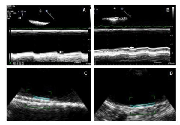
Figure 2.

Two-dimensional (2-D) semi-automated imaging of the aortic intima-media thickness. A 41 year old SLE patient with mid (A) and distal (B) aortic IMT of 0.72 mm (arrow) and 0.65 mm (arrow), respectively, by 2-D guided M-mode imaging. Corresponding IMT measurements by 2D-semi automated imaging were 0.75 mm (C) and 0.68 mm (D), respectively.
